
The ultrasound examination can be one of the most exciting, and nervous, events of your pregnancy. We help explain the process to help you feel more prepared and at ease going into the examination.
How does an ultrasound work?
The ultrasound transmits high-frequency sound waves that echo back and produce an image. Based on the timing of the reflected sound waves, the ultrasound machine can interpret the contours of the fetus, placenta, and umbilical cord.
Why are ultrasounds performed?
Typically ultrasound results can be exciting as they often can determine the sex of the baby, especially if it is a boy (boys have a more recognizable "structure"). But ultrasounds are performed for other reasons, including establishing the age of the fetus and accurately determining the due date, identifying fetal anomalies, determining if there is more than one fetus, determining the size and position of the fetus, seeing the baby's heartbeat, and assessing any pregnancy complications such as pain, bleeding, ectopic pregnancy, fibroids or tumors, or uterine structural problems.
When are ultrasounds typically performed?
It is possible to see the baby's heartbeat as early as six weeks into the pregnancy. Typically, the first ultrasound is performed between 12 and 16 weeks into the pregnancy. Additional ultrasounds may be performed to monitor any complications of the pregnancy.
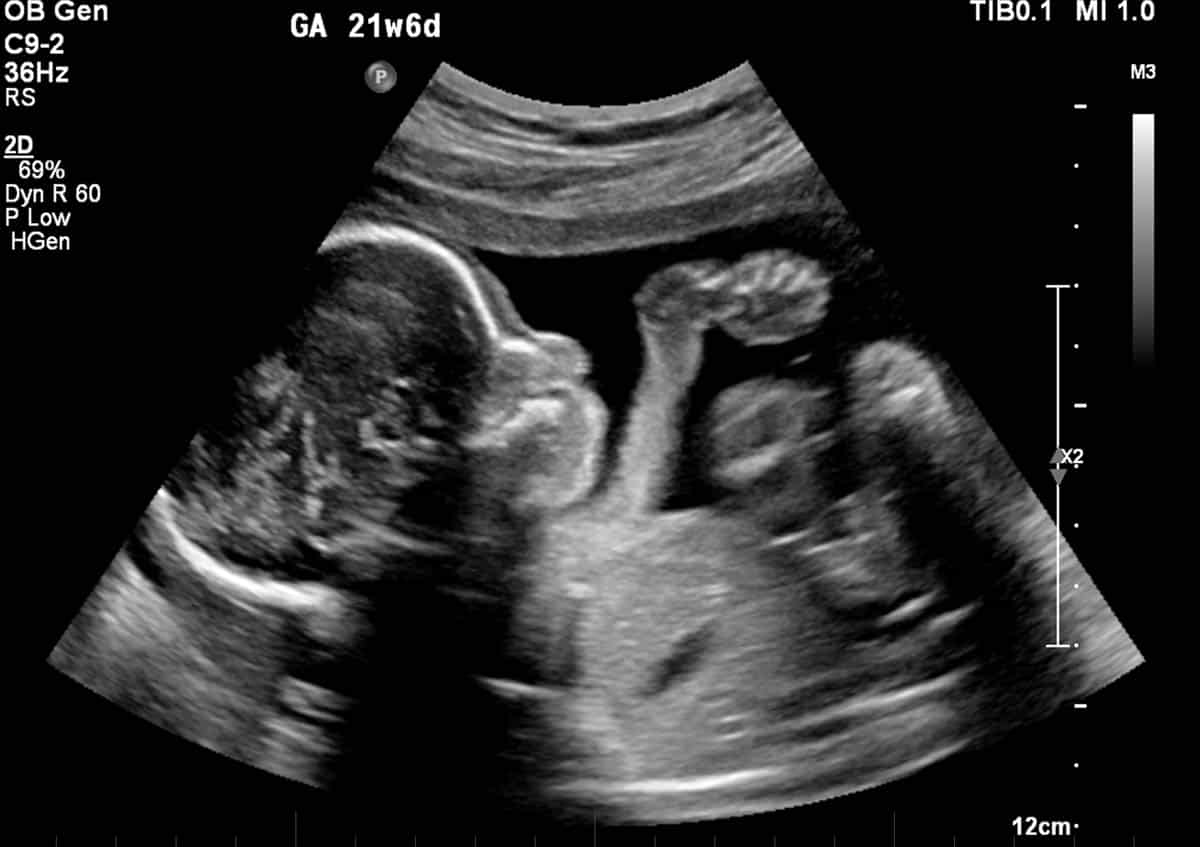
What are the different types of ultrasounds?
In addition to "traditional" ultrasounds, 3D and 4D ultrasounds are available. Traditional ultrasounds provide a grainy, black-and-white image that can be akin to interpreting Rorschach ink blot tests. Bones show up as white, tissues show up in gray, and fluid as black in the scan. Health insurance companies typically will cover traditional ultrasound screenings. 3D ultrasound screenings are provided in color, and provide a three-dimensional model of the fetus, which resembles a color photograph. 4D ultrasounds add time as the fourth dimension, providing a color video of the fetus moving around inside.
How do I prepare for the ultrasound exam?
Your doctor or medical practitioner will provide instructions for you. Typically, if you are less than 14 weeks along, you will be asked to arrive at the appointment with a full bladder, as the ultrasound waves travel better through the liquid. The ultrasound technician will squirt a clear gel on the abdomen and spread it around so that the ultrasound probe, or transducer, can slide around smoothly. Abdominal ultrasounds are the most common, but vaginal ultrasounds are also performed. In this technique, a wand-shaped probe is covered with a latex sheath and lubricant and placed into the vagina. You may be asked to bring in a blank videotape if you would like a copy of the ultrasound video, although the technician's voice is generally not added to the video for liability and confidentiality reasons.
Who performs the ultrasound exam?
Generally, a trained ultrasound technician will perform the examination. They identify the different body parts and are usually very good about showing the different parts to the parents. If complications are detected or your pregnancy is considered a high-risk pregnancy, a perinatologist or radiologist may perform the scan.
Note: Please also check your spam or junk email folder.